Introduction
If you've recently undergone a total knee replacement surgery, congratulations on taking this important step toward improved mobility and quality of life. As part of your post-surgical care, your orthopedic surgeon will order several X-rays to monitor your new knee's position and healing progress. These X-rays serve as vital tools for your medical team, but they can appear quite mysterious to patients who aren't familiar with medical imaging.
Dr. Debashish Chanda understands that seeing metal components where your natural knee joint once existed can be both fascinating and potentially concerning. This comprehensive guide aims to demystify total knee replacement X-rays, helping you understand what you're looking at, why these images are crucial for your recovery, and what your surgeon is evaluating when they examine these pictures.
What Is a Total Knee Replacement X-Ray?
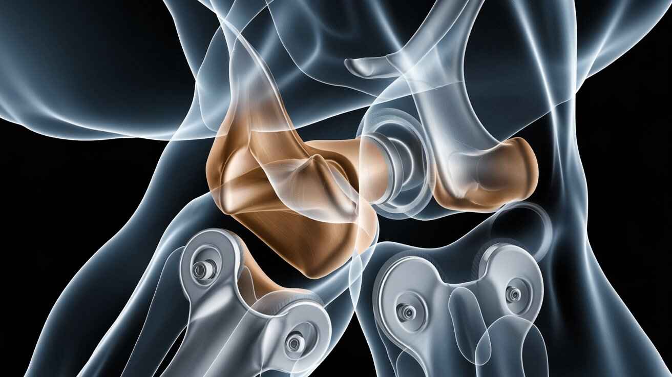
A total knee replacement X-ray is a specialized radiographic image that shows the artificial components of your new knee joint and how they interact with your existing bone structure. Unlike a regular knee X-ray that shows natural anatomy, your post-surgery X-rays will reveal the metal and plastic components of your prosthetic knee.
Components Visible on Your X-Ray
When you look at your total knee replacement X-ray, you'll notice several distinct components:
- Femoral Component: This metallic piece caps the end of your thigh bone (femur) and appears as a curved, bright white shape at the top of your knee joint.
- Tibial Component: This flat, metallic platform sits on top of your shin bone (tibia) and shows up as a bright white horizontal line below the femoral component.
- Polyethylene Insert: This plastic spacer between the metal components doesn't appear directly on X-rays but is visible as a gap between the femoral and tibial components.
- Patellar Component: If your kneecap (patella) was resurfaced during surgery, you might see a small button-like component on the front view of your knee.
- Your Natural Bones: The femur (thigh bone), tibia (shin bone), and fibula (smaller lower leg bone) appear as lighter gray structures surrounding the bright white metal components.
Why Are Post-Operative X-Rays Necessary?

Total knee replacement X-rays serve multiple crucial purposes throughout your recovery journey:
Immediate Post-Surgical Assessment
X-rays taken immediately after surgery allow Dr. Chanda to verify:
- Proper alignment of all components
- Correct positioning relative to your natural bone
- Appropriate spacing between components
- Absence of any immediate complications
Short-Term Follow-Up (6 Weeks to 6 Months)
Follow-up X-rays help monitor:
- Early signs of bone integration with the implant
- Maintenance of proper alignment as you begin using the knee
- Absence of subsidence (sinking) of components into bone
- No signs of early loosening or wear
Long-Term Monitoring (Annual or Biennial)
Regular X-rays in the years following surgery help detect:
- Long-term stability of the components
- Signs of wear in the polyethylene insert
- Potential loosening of the implant
- Changes in bone density around the implant (osteolysis)
- Proper function with your natural movement patterns
How to Read Your Own Total Knee Replacement X-Ray

While your surgeon is trained to evaluate subtle details on your X-ray, understanding some basics can help you have more informed conversations during your follow-up appointments.
Different Views and What They Show
Typically, your orthopedic team will take several different angles of your knee:
1. Anterior-Posterior (AP) View
- Taken from the front of your knee
- Shows the width of the components and side-to-side alignment
- Reveals how the femoral component sits on the tibial component
2. Lateral View
- Taken from the side of your knee
- Shows the position of components from front to back
- Reveals the position of your kneecap relative to the joint
3. Merchant or Sunrise View
- Taken with your knee bent and from above your kneecap
- Shows how your kneecap tracks within the groove of the femoral component
Normal Findings on a Total Knee Replacement X-Ray
Here's what Dr. Chanda looks for as signs that everything is proceeding normally:
- Proper Alignment: The components should form a straight line when looking at the leg from front to back
- Good Contact: The femoral component should sit evenly on the tibial component
- Appropriate Joint Space: The gap between components should be consistent
- No Radiolucent Lines: There shouldn't be dark lines between the implant and bone (which could indicate loosening)
- Bone Growth: Over time, you may see slight haziness around the implant edges where bone is growing into the implant surface
When to Be Concerned About Your X-Ray Results
While your surgeon will identify any issues, it's helpful to understand potential red flags that might require further attention:
Potential Abnormal Findings
- Component Misalignment: Components that appear tilted or at odd angles to each other
- Radiolucent Lines: Dark lines between the implant and bone that may indicate loosening
- Fractures: Breaks in the surrounding bone tissue
- Joint Effusion: Excessive fluid around the joint visible as increased soft tissue shadows
- Heterotopic Ossification: Abnormal bone formation in soft tissues around the implant
- Osteolysis: Loss of bone density around the implant components
Remember that not all abnormalities on an X-ray cause symptoms, and conversely, not all symptoms have visible causes on X-rays. This is why communication with Dr. Chanda about both your images and your physical symptoms is essential.
The Timeline of X-Rays After Total Knee Replacement
Understanding when X-rays will be taken helps you prepare for your follow-up care:
Immediate Post-Operative (Day 0-1)
Your first X-ray will likely be taken before you leave the hospital, sometimes even in the operating room. This serves as a baseline for future comparisons and confirms proper initial placement.
First Follow-Up (2-6 Weeks)
This X-ray helps assess early healing and ensures components remain properly positioned as you begin physical therapy and more movement.
Three to Six Month Follow-Up
These images evaluate your progress as you return to more normal activities and assess how your bone is adapting to the implant.
One-Year Mark
This critical milestone X-ray establishes your "new normal" and serves as a baseline for long-term monitoring.
Annual or Biennial Long-Term
Depending on your age, activity level, and specific implant, Dr. Chanda will recommend a schedule for ongoing monitoring, typically every 1-2 years.
How X-Rays Complement Your Physical Examination
X-rays are just one tool in evaluating your knee replacement's success. Your surgeon will always correlate imaging findings with:
- Range of motion measurements
- Stability testing
- Pain assessment
- Functional abilities
- Your reported symptoms
This comprehensive approach ensures that both objective imaging data and your actual experience inform your ongoing care plan.
Beyond X-Rays: Other Imaging That May Be Used
While X-rays remain the standard for routine monitoring, sometimes additional imaging may be necessary:
CT Scans
Computerized tomography provides detailed cross-sectional images that can help evaluate:
- Complex alignment issues
- Suspected fractures not visible on X-ray
- Detailed bone loss patterns
MRI With Metal Artifact Reduction
Though challenging with metal implants, specialized MRI techniques can sometimes help assess:
- Soft tissue conditions around the implant
- Suspected infection or inflammation
- Unexplained pain sources not visible on X-ray
Bone Scans or SPECT Imaging
These nuclear medicine techniques can help identify:
- Areas of increased bone turnover
- Potential loosening not yet visible on X-ray
- Infection versus aseptic loosening
How to Prepare for Your Follow-Up X-Ray Appointments
Making the most of your imaging appointments ensures you receive optimal care:
Before Your Appointment
- Wear comfortable clothing that can be easily removed or pulled up above your knee
- Bring previous imaging results if you're seeing a new provider
- Make a list of questions you have about your X-rays
- Note any symptoms or concerns to discuss while viewing your images
During Your Appointment
- Ask to see your images - most surgeons are happy to show you what they're looking at
- Don't hesitate to ask questions about what you see
- Request clarification of any medical terms you don't understand
Commonly Asked Questions About Total Knee Replacement X-Rays
How much radiation exposure occurs during knee X-rays?
Knee X-rays involve minimal radiation - approximately 0.001 mSv per image, which is less than one day of natural background radiation. The benefits of proper monitoring far outweigh this minimal exposure risk.
Can I request copies of my X-rays?
Absolutely! You have the right to request digital copies or films of your images. These can be helpful if you need to consult with other providers or if you relocate.
Why do I need repeated X-rays if I'm feeling fine?
Even when you're symptom-free, regular imaging helps detect potential issues before they cause problems. Early identification of subtle changes can prevent more serious complications later.
What if my X-ray shows an abnormality but I have no symptoms?
This scenario is exactly why regular imaging is valuable. Sometimes problems develop gradually without causing immediate symptoms. Your surgeon will assess whether an asymptomatic finding requires intervention or simply closer monitoring.
Are there alternatives to X-rays for monitoring my knee replacement?
While other imaging modalities exist, X-rays remain the gold standard for routine monitoring due to their clarity in showing the bone-implant interface, low radiation, accessibility, and cost-effectiveness.
Conclusion: Becoming an Active Participant in Your Knee Replacement Journey
Understanding your total knee replacement X-rays empowers you to take an active role in your recovery and long-term joint health. These images tell an important story about your new knee - how it's positioned, how it's integrating with your body, and how it's holding up over time.
By familiarizing yourself with what appears on your X-rays and what your surgeon is evaluating, you can ask more informed questions and better understand your overall progress. Remember that X-rays are just one component of your comprehensive care plan with Dr. Debashish Chanda, complementing your physical exam, functional assessment, and personal experience with your new knee.
If you have questions about your total knee replacement X-rays or would like to schedule a consultation with Dr. Chanda to discuss your knee replacement journey, please contact our office. Together, we can ensure your new knee serves you well for many years to come.
Disclaimer: This article is for informational purposes only and should not be considered medical advice. Always consult with your healthcare provider regarding questions about your specific medical conditions, treatments, and follow-up care.

Debashish Chanda
Admin